Advanced Imaging Engine for Exceptional Clarity
▸ Equipped with a newly developed imaging engine, the APV-T8 delivers high-definition, noise-free images with improved tissue differentiation — ensuring accurate diagnosis even in challenging scanning conditions.
Specialized Cardiac and Abdominal Applications
▸ Supports advanced cardiac imaging functions, including Tissue Doppler Imaging (TDI), Strain Rate Imaging (SRI), and Anatomic M-Mode. Enables precise evaluation of heart structure and blood flow in animals of all sizes.
Real-Time Elastography and IMT Measurement
▸ Freehand elastography visualizes tissue stiffness in real time, ideal for breast, thyroid, and musculoskeletal analysis. Integrated IMT (Intima-Media Thickness) measurement assists in vascular and cardiac assessments.
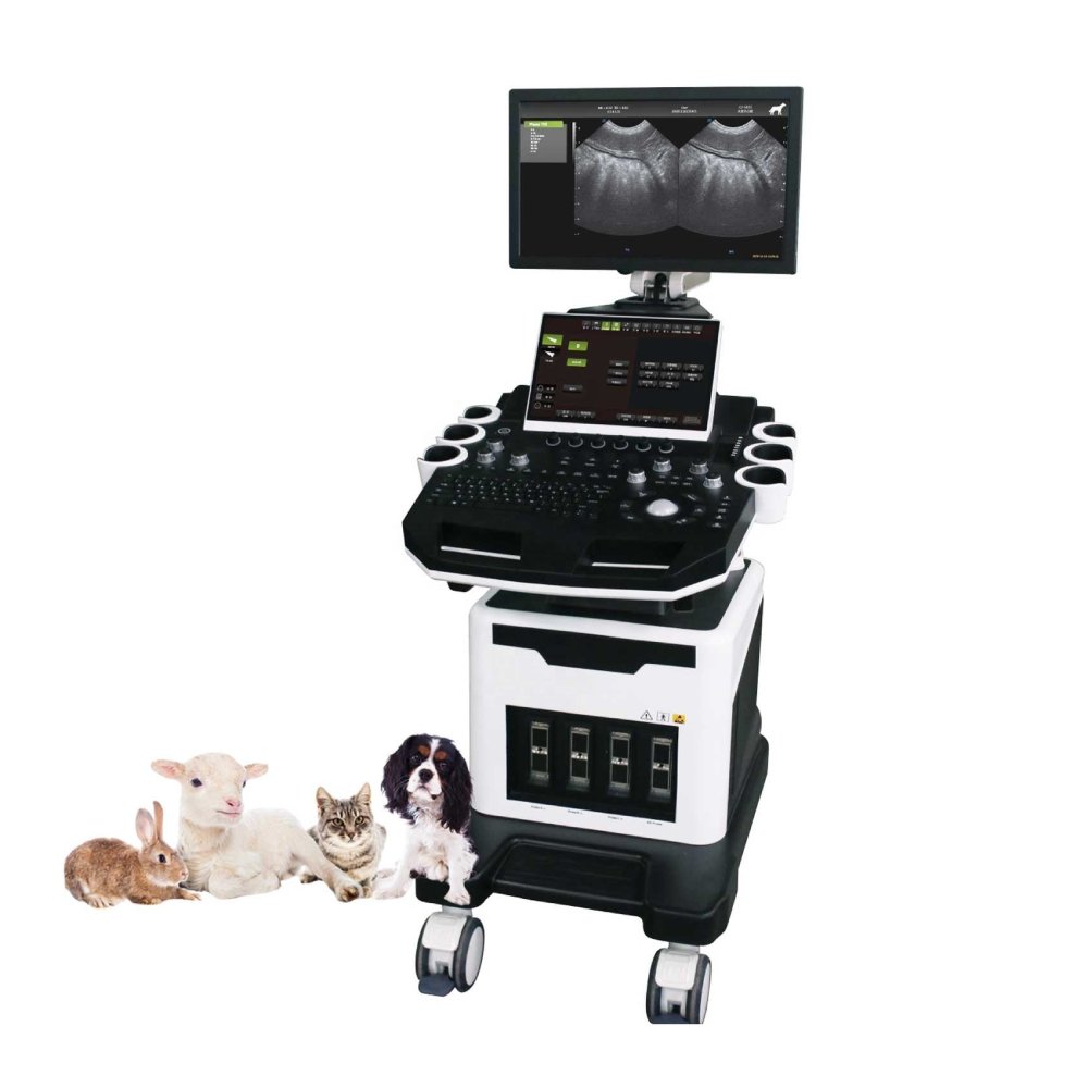
Versatile Use Across Multiple Species
▸ Designed for bovine, equine, canine, feline, ovine, and other domestic or wild animals. Suitable for clinical, research, and farm applications, including abdomen, thorax, tendon, eye, and reproductive examinations.
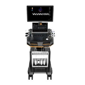
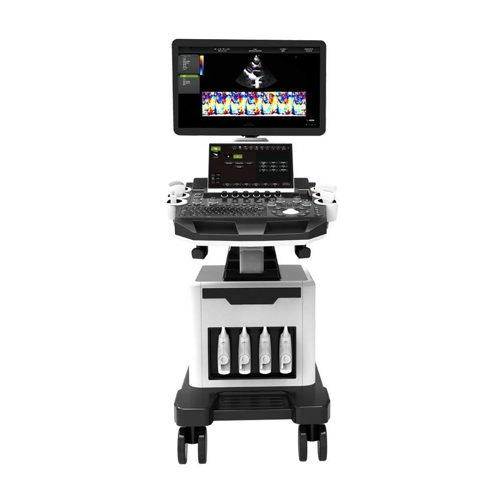
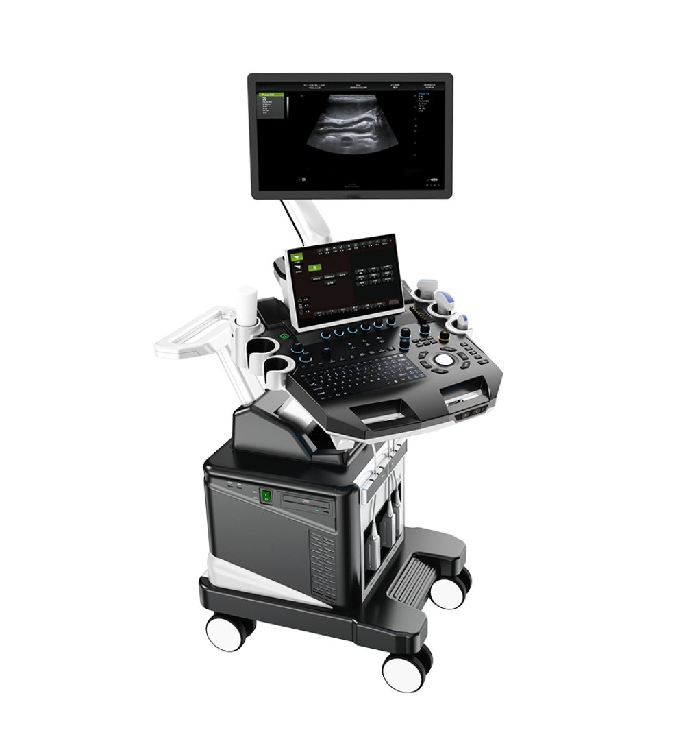
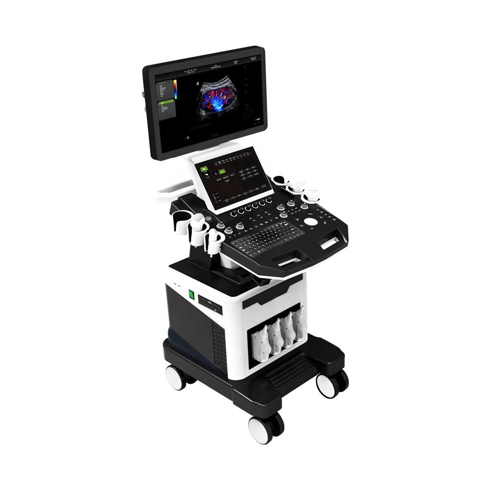
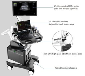
Dual-Screen Ergonomic Design
▸ Features a 21.5-inch medical display and a 13.3-inch touch screen for efficient workflow and easy operation. The newly designed console provides a comfortable and intuitive scanning experience.
Multi-Probe Connectivity for Comprehensive Scanning
▸ Four active probe ports allow instant switching between probes for different diagnostic needs — from convex and micro-convex to linear and rectal probes.
User-Friendly, Multi-Language System
▸ Supports multiple languages, including EN, FR, DE, RU, ES, PT, AR, VN, and ID — making it easy to use for global veterinary professionals and distributors.
Built for Stability, Mobility, and Long-Term Use
▸ Equipped with 4 lockable wheels, front/rear handles, and durable construction, ensuring safe movement and long-lasting reliability in clinical environments.
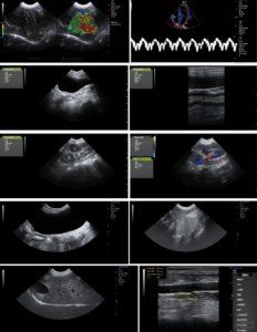
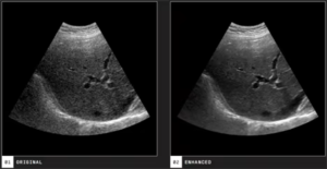
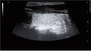
Speckle Noise Removal Technology
→ By analyzing ultrasound data across multiple spatial dimensions, this technology intelligently suppresses speckle noise while preserving fine tissue details. The result is exceptionally clear imaging that allows veterinarians to distinguish subtle anatomical structures and make faster, more confident diagnostic decisions.
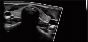
Real-time Wide-field Imaging
→ Expands the scanning field to capture large or complex lesions in real time. With picture-in-picture zoom, adaptive cropping, and motion stabilization, it ensures smooth, detailed visualization of large animals or extensive tissue regions, reducing the need for repeated repositioning during examination.
Omni-directional Adjustable M-Mode
→ This enhanced M-mode enables angle correction for more precise evaluation of cardiac structures. It captures accurate chamber dimensions and wall motion, even when the heart’s position makes scanning difficult—providing dependable results for comprehensive cardiac assessment.
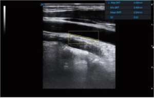
Automatic IMT Measurement
→ Automatically identifies and measures the intima-media thickness of blood vessels, a key indicator of cardiovascular health. The system optimizes measurement angles to deliver consistent and accurate vascular assessments, enabling early detection of potential cardiac risks in animals.
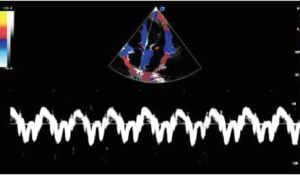
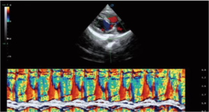
Tissue Doppler Imaging (TDI)
→ Measures the speed and direction of myocardial motion to provide a quantitative view of heart performance. TDI offers detailed insight into local myocardial function, helping veterinarians identify regional abnormalities and assess diastolic efficiency with precision.
Trapezoidal Imaging
→ Transforms linear probe data into an extended trapezoidal field of view without compromising resolution. This function enhances visibility for musculoskeletal and tendon examinations, especially in equine and small-animal diagnostics, where a wider image field is essential.
| Category | Specification |
| Display | 21.5-inch medical display, 13.3-inch touch screen for intuitive operation |
| Display Modes | B, BB, 4B, M, BM; Color Doppler: B/C, B/C/M, B/POWER, B/C/PW |
| Imaging Functions | IMT (Intima-Media Thickness) measurement,
Coherent Contrast Imaging (CCI), Real-time B/C/D synchronous imaging, Power Doppler (PDI), Direct Power Doppler (DPDI), Tissue Doppler Imaging (TDI), Strain Rate Imaging (SRI), Panoramic imaging, Deflection imaging, Trapezoidal imaging, Freehand 3D imaging |
| System | Windows 7 |
| Obstetric & Species Measurement | Bovine (BTD, BUD),
Equine (EGSD, ESD), Swine (HLA, SLA), Ovine (SCRL), Canine (HD, BD, GSD, CRL), Feline (HD, BD) |
| Interfaces | HDMI, RJ-45, USB ×4, Grounding Wire, DVD-RW |
| Weight | 90 kg |
| Size | 607 × 976 × 1458 mm |
| Languages Supported | CN, EN, VI, DE, FR, ES, RU, AR, PT, ID |
| Optional Probes | Suitable Animals | Typical Use | Application Scenarios |
| Convex Probe | Pigs, sheep, and large dogs | Abdominal organ scans, pregnancy diagnosis | Livestock farms, breeding centers, and large animal clinics |
| Micro-Convex Probe | Cats, small & medium-sized dogs | Abdominal organ and pregnancy scans | Pet hospitals, veterinary clinics, and research institutions |
| Linear Probe | Small animals | Abdominal organs, musculoskeletal, and subcutaneous tissues | Pet hospitals, veterinary practices |
| Rectal Probe | Cattle, horses | Pregnancy checks, reproductive system exams | Cattle ranches, horse farms, and reproduction management |
| Back Fat Probe | Pigs, cattle | Measuring back fat thickness and muscle condition | Livestock farms, feedlots, meat production facilities |
| Phased Array Probe | dogs, cats, livestock | Cardiac diagnosis | Veterinary clinics, research institutions, and animal hospitals |
| 4D Probe | Bovine, equine, ovine, canine, feline | Real-time 4D fetal imaging | Reproductive centers, veterinary hospitals, breeding farms |