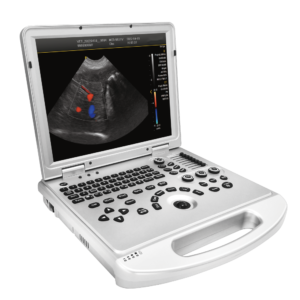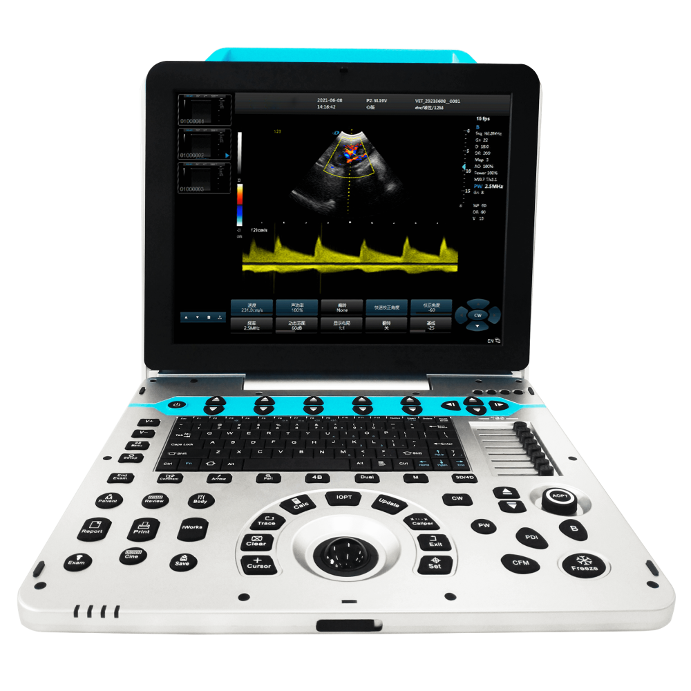
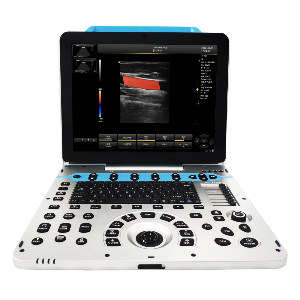
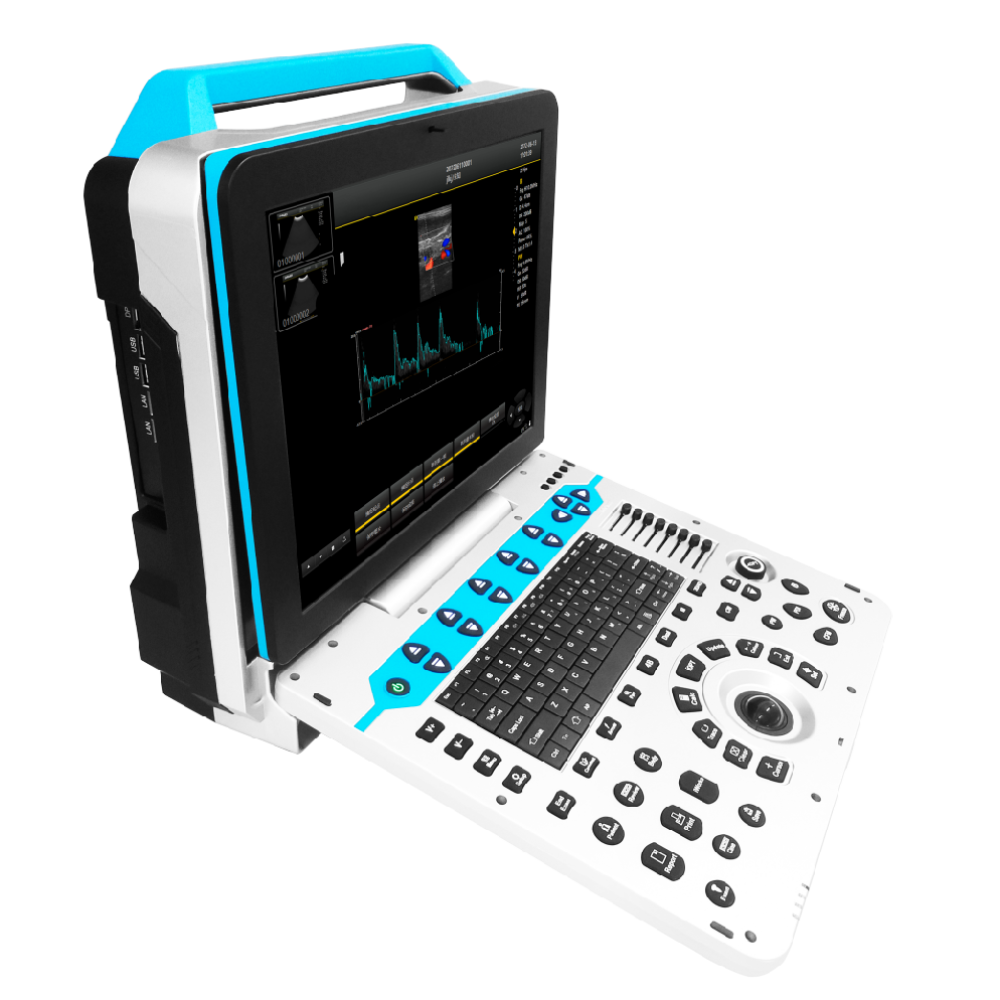
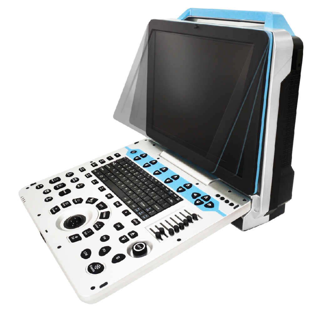
This portable veterinary ultrasound system is designed for veterinary clinics, breeding centres and mobile practitioners requiring reliable imaging performance for both companion animals and livestock applications.
With advanced imaging technologies, flexible probe options and dedicated veterinary measurement packages, it supports routine diagnostic examinations, reproductive assessment and clinical imaging across a wide range of animal species.
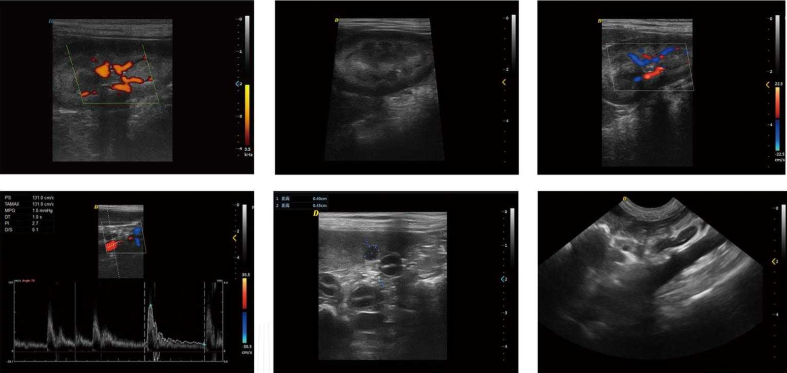
Example ultrasound images demonstrating imaging performance in veterinary examinations.
| Display | 15-inch high-resolution medical screen |
| Imaging Modes | B, BB, 4B, M, BM |
| Color Doppler Modes | B/C, B/C/M, B/POWER, B/C/PW |
| Advanced Imaging | Coherent Contrast Imaging (CCI), Power Doppler Imaging (PDI), Direct Power Doppler Imaging (DPDI), Tissue Doppler Imaging (TDI), Strain Rate Imaging (SRI) |
| Real-Time Imaging | B/C/D triple synchronous imaging |
| Veterinary Measurement Packages | Bovine, Equine, Swine, Ovine, Canine and Feline measurement packages |
| System | Windows 10 |
| Storage | 4G + 128G high-speed PC platform |
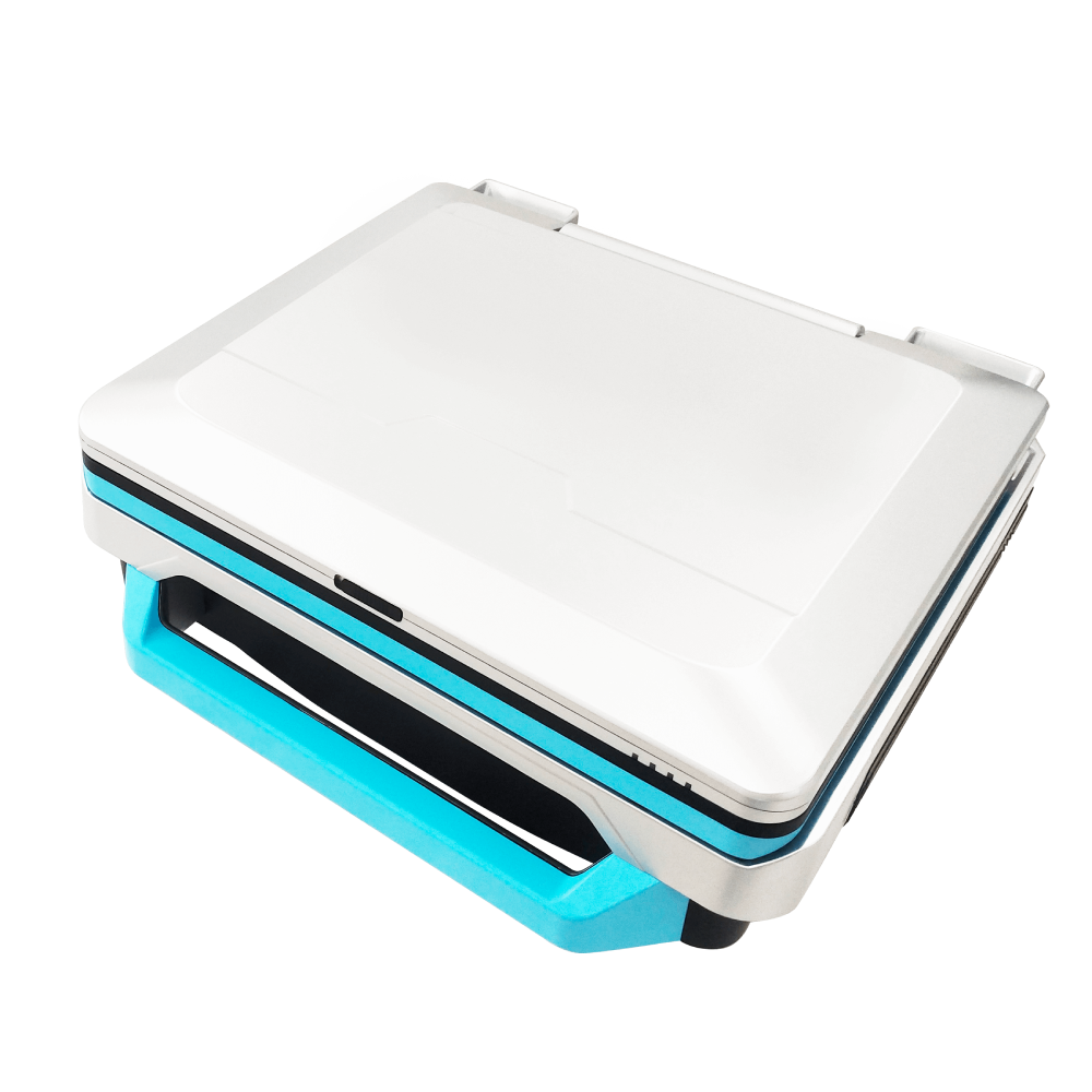
| Weight | 8.6 kg |
| Dimensions | 376.7 × 166.7 × 386.4 mm |
| Interfaces | 4 × USB, 1 × Audio, 2 × RJ-45, 1 × HDMI, 1 × DP |